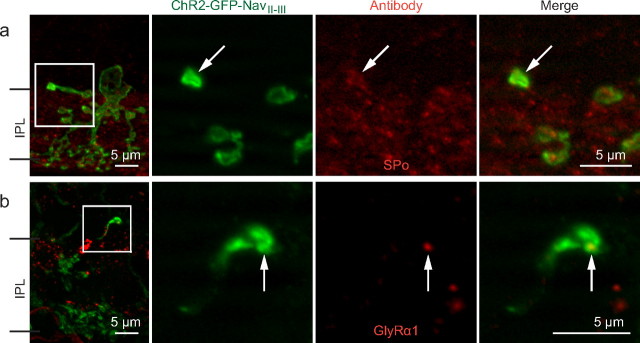
Figure 3.
IHC characterization of lobular appendage features in AII processes. a, b, The first panels are z-stack projections (20× objective; 1 μm z-sections), and three subsequent panels are single-plane magnifications (63× objective, 0.6 μm z-sections) of the corresponding boxed areas showing an AII process terminal colocalizing with SPo immunostaining (a) and an AII process colocalizing with a GlyRα1 punctum (b).

