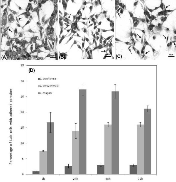
Figure 1.

Analysis of promastigote adhesion to Lulo cells. After interaction between Lulo cells and promastigotes, the samples were stained with Giemsa for analysis. In the figure, the arrows are indicating the promastigotes adhered to Lulo cells at 48 h for Leishmania (V.) braziliensis (A), Leishmania (L.) amazonensis (B) and 24 h for Leishmania (L.) chagasi (C). Quantitative data (D) of the interaction were assessed at different times (2 h; 24 h; 48 h; 72 h) of incubation. Data are expressed in percentile values (%) and represent average and standard deviation of five independent experiments.
