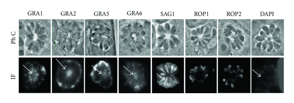
Figure 3.

Localization of GRA proteins in the RB. MDCK cells infected with RH tachyzoites were incubated with antibodies directed against the dense granule proteins GRA1, GRA2, GRA5, and GRA6, rhoptry proteins (ROP1, 2), or membrane protein SAG1. GRA proteins marked the RB (arrows), while the stain with nuclear marker, DAPI, showed a weak staining of RB. Immunofluorescence micrographs (IF) are shown with their respective phase contrast microscopy images (PhC).
