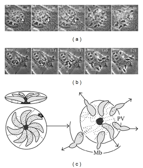
Figure 8.

During exteriorization of tachyzoites, the residual body remains inside the PV. (a) Egress of RH tachyzoites arranged as “rosettes” from infected MDCK cells was induced by 0.1 μm ionomycin and recorded by time lapse videomicroscopy. Externalizing parasites are indicated by the arrows. Arrowheads show the constriction of the tachyzoites passing through the host plasma membrane. The asterisk indicates remnants of the RB after the egress of parasites from a rosette. (b) The exteriorization of tachyzoites of ΔGRA2 strain was slightly slower than the RH strain. Several ΔGRA2 tachyzoites remained trapped in the cytoplasm or the nucleus (asterisks). Numbers in the upper right corners indicate the timing in seconds. N: nucleus of the host cell. Bars = 5 μm. (c) represents the individual exteriorization routes followed by tachyzoites organized in rosettes.
