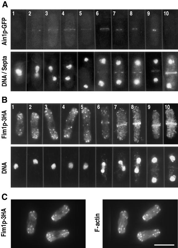
Figure 3.
Localization of Ain1p and Fim1p during the cell cycle. In A and B, upper and lower panels show the same cells, and individual cells are numbered for reference in the text; in C, the two panels show the same cells. (A) Localization of Ain1p to the medial ring. Cells expressing Ain1p-GFP (strain JW46) were grown in EMM medium at 25°C and stained with Calcofluor and bisBenzimide to allow visualization of septa and DNA. (B and C) Localization of Fim1p to actin patches and the medial ring. Cells expressing Fim1p-3HA (strain JW106) were grown in EMM at 30°C, fixed, and double-stained either with HA-specific antibody and bisBenzimide (B) or with HA-specific antibody and rhodamine-phalloidin (C). Bar, 10 μm.

