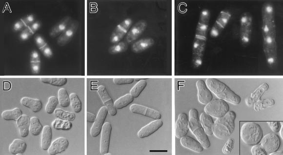
Figure 5.
Morphological abnormalities of ain1 deletion and fim1 deletion cells. (A–C) Cells were double-stained with Calcofluor and bisBenzimide to visualize septa and DNA. (D–F) Cells were observed directly in culture medium by DIC microscopy. (A) ain1-Δ1 cells (strain JW45) grown in EMM medium at 25°C. (B and C) Wild-type strain 972 (B) and strain JW45 (C) grown in EMM + 1 M KCl medium at 18°C for 40 h. (D) fim1-Δ1 cells (strain JW142) grown in EMM at 25°C. (E and F) Strains 972 (E) and JW142 (F) grown for 5 h (20 h for the cells in the inset) after a shift from 25 to 36°C in EMM. Bar, 10 μm.

