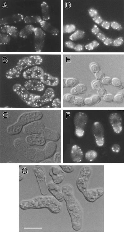
Figure 9.
(A–F) Localization of Fim1p-GFP during mating (A), sporulation (B and D), and spore germination (F). (C and E) DIC images of the cells shown in B and D, respectively. fim1+-GFP strains JW109 and JW117 were crossed on an SPA plate at 25°C, and samples taken after 12 h (A), 24 h (B and C), and 50 h (D and E) were examined after resuspension in liquid SPA medium. After 60 h, the mature asci were digested with Glusulase (NEN, Boston, MA) to release spores, which were then incubated in liquid EMM medium at 25°C and examined after 12 h (F). (G) Defective sporulation in fim1 deletion cells. fim1-Δ1 strains JW144 and JW142 were crossed on an SPA plate at 25°C and examined by DIC after 50 h. Bar, 10 μm.

