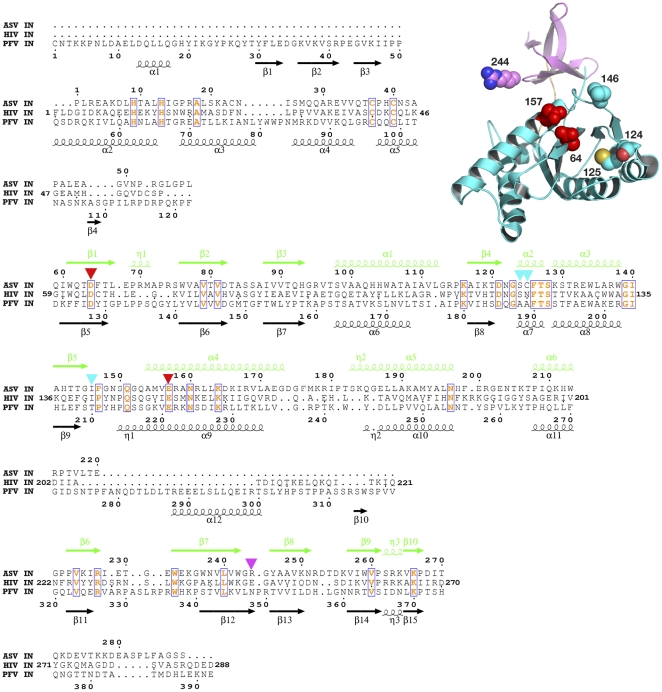
Figure 2. Structure-based sequence alignment of full-length ASV, HIV-1, and PFV IN proteins.
ASV IN numbering is shown above the sequences and the structural elements are marked in green; PFV IN numbering and structural elements (black) are shown below. Numbering for HIV-1 IN is shown at the beginning and end of the lines only. The conserved amino acids, including the catalytic ASV IN residues Asp121 and Glu157, are red and boxed. Triangles mark residues that were changed to cysteines in ASV IN: red for the amino acids in the active site, cyan for other residues in the CCD, and magenta for the amino acids in the CTD. The structure of the ASV IN CCD and CTD (PDB code 1COM) with the location of the introduced cysteines is shown in the upper right corner, with the colors corresponding to the scheme described above.

