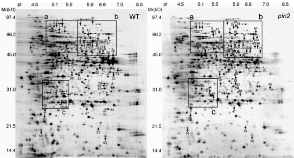
Figure 3.

Images of 2-D gels of proteins extracted from Arabidopsis wild type (WT) and pin2 mutant (pin2) roots tips exposed to either clinostat rotation or hypergravity. The proteins were visualized by silver staining. The arrows indicate 54 protein spots in the WT (left panel) and 64 protein spots in the pin2 mutant (right panel) root tips that changed in abundance and/or position after at least one treatment of vertical clinorotation, horizontal clinorotation, and hyper gravity centrifugation. The sum number of the protein spots in both gels is 88 including 30 overlaps. The background gel was of a stationary control. The ranges of pI and molecular masses (kDa) are indicated. The framed regions a, b, and c are detailed in Figure 4.
