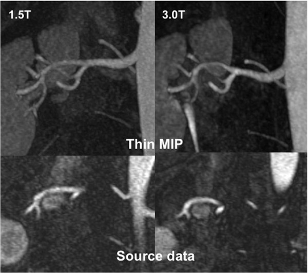
Figure 2.
Source data as well as thin MIP reconstructions of renal MRAs are provided, acquired in the same patient at 1.5 and 3 T. Although the renal vasculature is clearly depicted at 1.5 T, the SNR and CNR gains at 3 T provide even more homogeneous vessel signal, improving visualization of smaller subsegmental branches.

