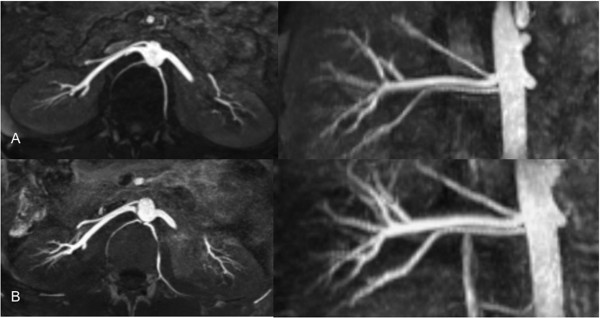
Figure 5.
Axial and coronal reformats of native TrueFISP renal MRAs are shown, acquired at 1.5 (A) and 3 T (B). These techniques are based on the inflow of arterial blood into the imaging plane, which is made visible by a SSFP-type of read-out scheme which is available in numerous vendor specific variations. Whereas, NCE-MRA appears to be a suitable screening tool for renal artery stenosis detection, accurate stenosis grading might not always be feasible. Courtesy of PD Dr. Blondin, University of Dusseldorf.

