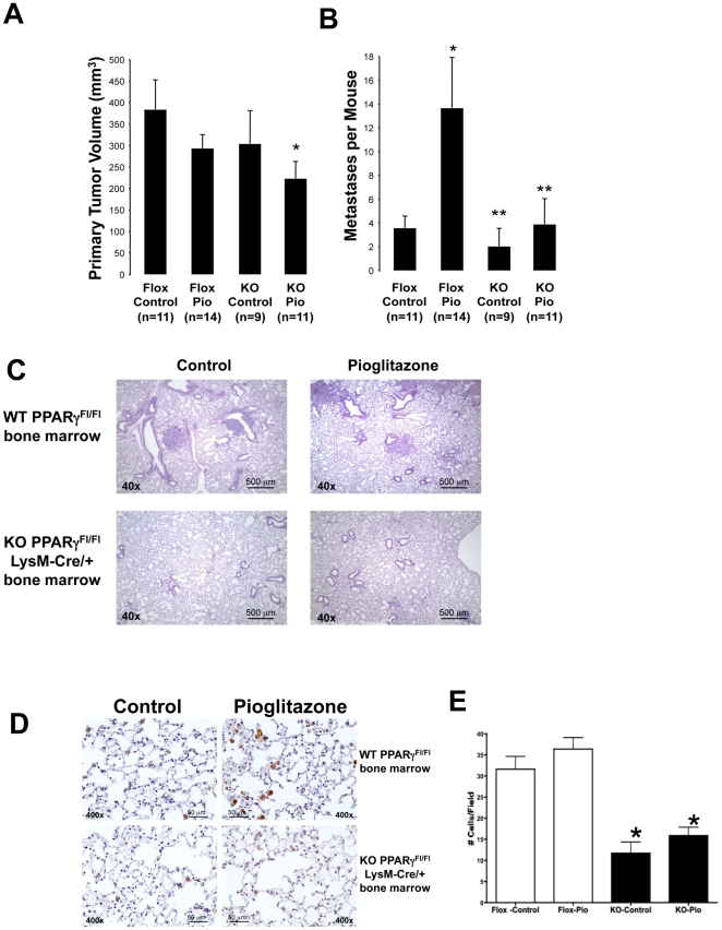
Figure 4. Targeted deletion of PPARγin Macrophages inhibits lung cancer metastasis in a subcutaneous flank model of murine non-small cell lung cancer.
Following lethal irradiation, WT C57BL/6 mice received bone marrow from either PPARγ-Macneg (KO) or PPARγflox/flox (Flox) donor mice as described in the “Methods” section. After 5 weeks recovery to allow engraftment, mice were placed on either pioglitazone-containing chow or control chow for 1 week prior to tumor cell implantation and throughout the course of the experiment. Animals were then injected with 105 CMT/167-luc cells subcutaneously. Animals were imaged by bioluminescence, and sacrificed 4 weeks after cancer cell inoculation. A. Primary tumor volumes in all the mice were measured using digital calipers. Data are means and s.e.m. of counts from 9–14 animals in each group. *P<0.05 vs Flox Control. B. Incidence of lung metastasis was quantitated by examination under a dissecting microscope. Tumors were counted by two independent blinded observers. Data are means and s.e.m. of counts from 9–14 animals in each group. Pioglitazone increased incidence of metastasis in WT mice, but not in mice receiving PPARγ-Macneg bone marrow. *P<0.05 vs Flox Control. **P<0.05 vs Flox Pio. C. Representative histology is shown for lung metastases from all 4 groups of animals. D. Tumor-bearing lung sections from WT or PPARγ-Macneg mice were immunohistochemically stained for arginase I (brown reaction color). Representative images are shown of lungs from all 4 groups of animals. E. The number of arginase I-positive cells was counted by two independent blinded observers. Data are means and s.e.m. of counts from 9–14 animals in each group with one section per animal and 4 random fields per slide. The number of arginase I-positive cells was significantly decreased in PPARγ-Macneg mice, both under control conditions and in the presence of pioglitazone. *P<0.05 vs Flox Control.

