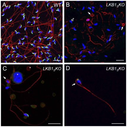
Figure 2. Fluorescence microscopy images of mature spermatozoa.
Spermatozoa were taken from the cauda epididymis of wild-type (A) and LKB1SKO mice (B–D), and visualised by immunofluorescence. Tails were stained with an anti-tubulin antibody (red), acrosomes with an anti-acrosomal monoclonal antibody (green) and nuclei with DAPI (blue). Sperm from LKB1SKO mice frequently show coiled tails (filled white arrows). In (D), the tail has formed a ‘lasso’-type structure around an abnormally shaped nucleus. Sperm nuclei from LKB1SKO mice often lack acrososmes (open white arrow in B) as shown by the lack of green fluorescence at the anterior nuclear surface. Abnormal cellular debris is visible in LKB1SKO samples, as indicated by asterisks in (C). Slides were viewed on a Leica TCS SP1 confocal microscope. Images are representative of at least three mice (Scale bar = 20 µM).

