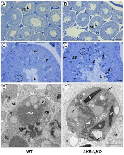
Figure 4. Testis histology.
Cross-sections of seminiferous tubules from wild-type (left-hand panel, A and C) and LKB1SKO (right-hand panel, B and D) mice. The seminiferous epithelium (SE), which is made up of sertoli cells and developing germ cells, is indicated. This surrounds the lumen (L) of the tubules. (C) and (D) show sections from tubules at approximately stage V. Circles have been used to show the nuclei of elongating spermatids. Spermatogonia (open curved arrow), spermatocytes (closed curved arrow) and round spermatids (open arrow) are also indicated. A number of dense, round bodies of varying sizes can be seen around the lumens from LKB1SKO mice. Examples of these are indicated in (D) (closed black arrows). Representative images are shown (A and B, scale bar = 100 µm; C and D, scale bar = 20 µm). (E) and (F) show TEM images of a residual body (RB) within the seminiferous epithelium (SE) from a wild-type mouse (E), and an abnormal cytoplasmic body from a LKB1SKO mouse (F). Normal residual often contain such structures as vacuoles (V), RNA and mitochondria (Mt). The abnormal cytoplasmic bodies seen in (F) are similar to residual bodies but often contain at least one condensed spermatid nuclei (N) and several cross sections of flagella (F) as indicated. Detached acrosomes, identified as the deeply staining crescent shape structures close to the anterior nuclear surface, are indicated with an arrow. Granular material is also indicated (G), (E and F, scale bar = 2 µm).

