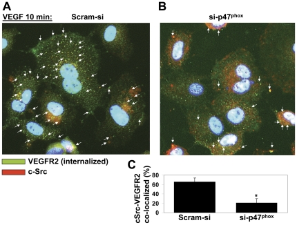
Figure 4. VEGF induces subcellular co-localization of c-Src and internalized VEGFR-2 in an ROS-dependent manner.
HCAEC transfected with control (Scram-si) (A) or si-p47phox (B) were immunofluorescently double labeled for internalized VEGFR-2 (green) and c-Src (red). VEGFR-2 on HCAEC was labeled with single chain E-tagged antibody (scFvA7, Fitzerald) as described in Materials and methods. After incubation with VEGF (50 ng/ml for 10 min), in order to remove the antibody from the cell surface, cells were placed on ice and acid washed. In permeabilized and fixed HCAEC, VEGFR-2 was detected with an AlexaFluor488-conjugated secondary antibody and is shown in green. c-Src was labeled with AlexaFluor647-conjugated secondary antibody (red) and nuclei with DAPI (blue). (B) Bar graphs show image analysis for colocalization events using the NIH Image J plugin (as described in Materials and methods). The graphs present the number of colocalization events normalized for the number of total VEGFR-2–positive immunofluorescence signals. Values are the mean of three experiments ± S.E.M., each containing numbers obtained from five random fields. *p<0.05 was considered statistically significant.

