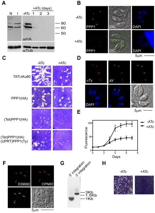
Figure 7. PPP1 is essential for parasites growth.
(A) Western blot analysis was of proteins extracts from PPP1(HA) and (Tet)PPP1(HA) using anti HA antibody. Note comparable levels of PPP1 when expressed from the native promoter (N) or the inducible T7S4 promoter (I). Protein levels decline swiftly upon ATc treatment, numbers indicate days of culture in 0.5 µg/ml ATc. Tubulin (lower panel) serves as a loading control. (B) Fluorescence microscopy analysis of (Tet)PPP1(HA parasites using anti-HA antibody grown in the presence and absence of ATc. (C) Plaque assays performed in the absence (-) or presence (+) of ATc. Parasite line name is indicated to the left. (D) Fluorescence microscopy of parasites expressing a second copy of PPP1, which is labeled using an anti-Ty antibody (red). The apicoplast marker used is the endogenously tagged ACP-YFP (AY, green). Scale bar = 5 µm. (E) Fluorescence growth assays show growth deficiency in ATc treated (Tet)PPP1(HA). Circles, -ATc; squares, +ATc. Each data point represents the mean of six wells, and the error bar gives the standard deviation. (F) Fluorescence microscopic analysis of endogenously tagged 039680 using anti-HA (green) and the luminal marker CPN60 (red) show that 039680 is likely a luminal apicoplast protein. (G) PCR analysis of the promoter replacement clone of 039680 with diagnostic primer sets is consistent with locus modification. The size of amplicons matches the prediction for integration at both the 5' and the 3' insertion site (900 and 1800 bp respectively). (H) Plaque assays performed with the promoter replaced 039680 cell line in the presence or absence ATc show that this gene is required for growth.

