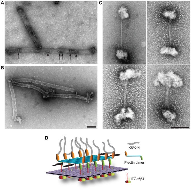
Figure 8. RD polymer formation and model highlighting lateral RD association as a novel HD-stabilizing force.
(A,B) Electron microscopy of polymeric RD structures. pHLH20/wt-encoded recombinant versions of plectin's RD (Figure 6A) were incubated in 50 mM sodium phosphate, pH 7.4, 300 mM NaCl, 170 mM imidazole, and 1 mM PMSF for 1 hour at 37°C, before being processed for uranyl acetate staining and electron microscopy. Note that the slightly larger diameter of flat sheet/envelope-like structures visualized in (A) would correspond to that expected for collapsed (flat) tube-like structures visualized in (B). Constrictions (A, arrows) of sheet-like structures may represent transition states between flat (collapsed) and tube-like structures. Bar, 100 nm. (C) Electron microscopy of oligomeric dumbbell-like structures assembled from intact (full-length) plectin molecules. Plectin samples purified from rat glioma C6 cells in 7 M urea were dialysed into 2 mM Tris-HCl, pH 8.0, 5 mM β-mercaptoethanol, and 3 mM PMSF at 4°C overnight. Samples were then incubated in the presence of 10 mM CaCl2 at 37°C for 1 hour, and thereafter processed for negative staining electron microscopy. Note correlation between increasing sizes of globular end domains and stalk diameters of the four specimens shown. Bar, 100 nm. (D) Schematic model depicting the major protein-protein interactions that provide the mechanical stability of HDs. Interaction of P1a dimers with ITGβ4 and K5/K14 provides a vertical force component, whereas the ITGβ4-induced lateral association of multiple dimeric P1a molecules (in an anti-parallel fashion via their RDs) could generate an additional horizontal force component (arrows), parallel to the plasma membrane (violet sheet). Note that individual proteins are not drawn to scale and BPAG1e and BPAG2 are not depicted. In contrast to classical models of HD protein organization, the sheet-like association of plectin molecules in our model is better suited to incorporate the dimensions of plectin molecules (>230 nm) within the 50–60 nm thick HD inner plate.

