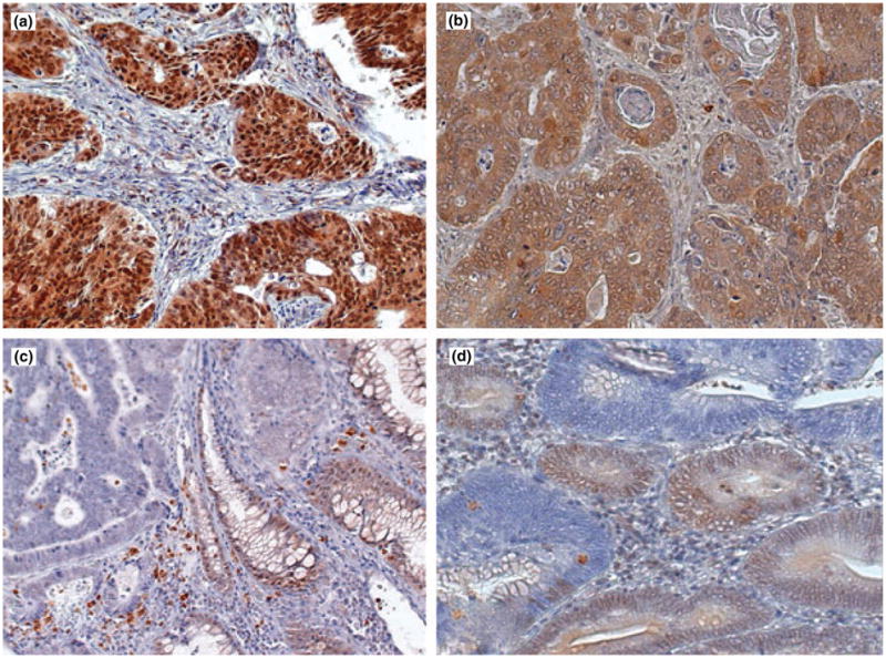
Figure 1.

Immunohistochemical staining for SMAD4 performed using mAb B-8 in formalin-fixed paraffin-embedded sections of colorectal carcinomas. (a) Tumour showing strong expression of SMAD4 protein in the nucleus of tumour cells. (b) Tumour showing strong diffuse staining localized to cytoplasm with weaker nuclear staining. (c) Adenocarcinoma with loss of SMAD4 protein in tumour cells. Staining is present in adjacent normal colonic mucosa (arrow). (d) Representative section of tumour with focal positive and negative staining suggesting clonal differences in SMAD4 expression. Original magnification ×45.
