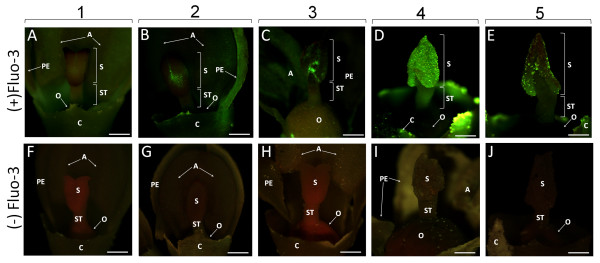
Figure 4.
Detection of Ca2+ by Fluo-3 AM in the pistils during olive flower development. Images were obtained using a stereomicroscope under blue light (488 nm). Microphotographs in the upper row show the buds/flowers taken from injected inflorescences [(+) Fluo-3], whereas the lower row shows control buds/flowers [(-) Fluo-3] from each corresponding developmental stage. (A) Green flower bud (stage 1): practically no labelling is present in the stigma. (B) White flower bud (stage 2): the labelling appears in some areas of the stigmatic surface. (C) Flower with turgid anthers (stage 3): well-distinguishable green fluorescence is located in the outer part of the stigma. (D) Flower with dehiscent anthers (stage 4): strong labelling is distributed throughout the stigmatic surface. Green fluorescence is also emitted from the stylar tissues. (E) Flower without sepals and petals (stage 5): the labelling is limited to small areas of the stigmatic surface. (F-J) Controls of the examined developmental stages (1-5). No green fluorescence can be detected in any analyzed stage. A - anthers, C - calyx, O - ovary, PE - petals, S - stigma, ST - style. Bars = 0.5 mm.

