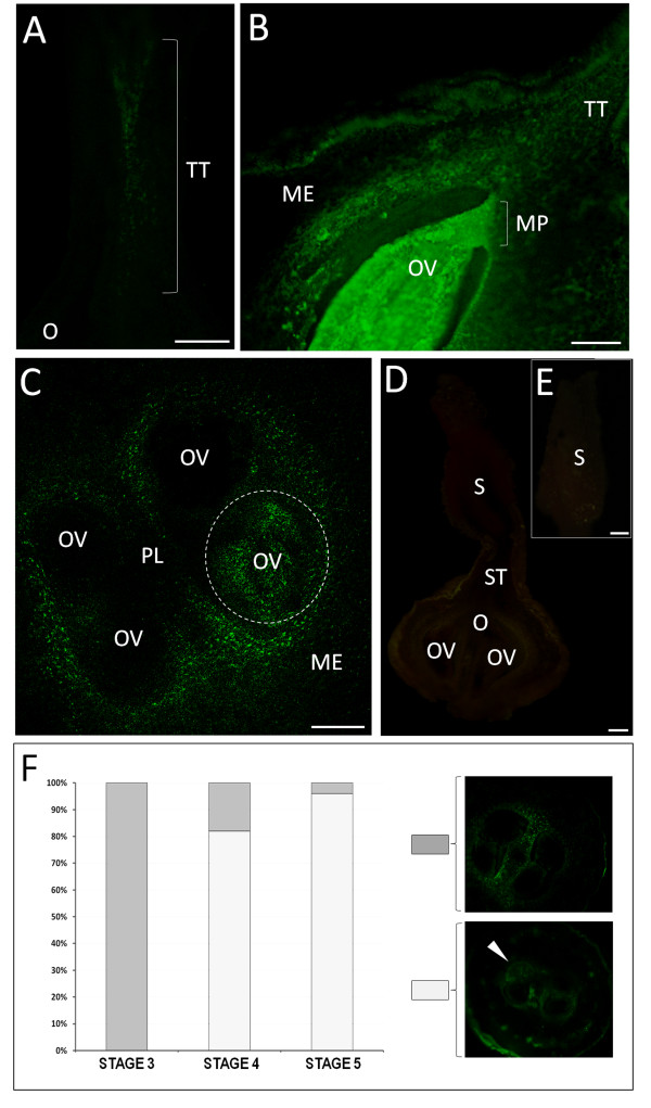
Figure 6.
Ca2+ detection in the internal parts of the pistil from flowers with dehiscent anthers. (A) In the longitudinally cut style, accumulation of green fluorescence is present in the area of the transmitting tract. (B) In the lower style and ovary, the labelling is located in the transmitting tract and around the loculus; stronger green fluorescence is localized in the whole area of the ovule, beginning from the micropylar region. (C) Transversal section of the ovary. Intense green fluorescence is visible in the areas directly surrounding 2 loculi and only in 1 of the 4 ovules present in the ovary (area marked with the dashed line). The remaining ovules show no signal. (D) Control reaction. In a longitudinally cut pistil that is not injected with Fluo-3, no green fluorescence can be detected in any part of the pistil. (E) Stigma of the control pistil. No green fluorescence is present in the papillae cells or in the attached pollen grains. ME - mesocarp, MP - micropylar region, O - ovary, OV - ovule, PG - pollen grain, PL - placenta, S - stigma, ST - style, TT - transmitting tract. Bars = 100 μm. (F) Graph comparing the percentage of ovaries where none of the ovules showed labelling with those where specific accumulation of Ca2+ only in 1 of the 4 ovules at stages 3, 4, and 5 was indicated.

