Figure 4.
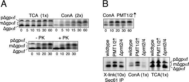
O-mannosylation protects Δgpαf from export to the cytosol. (A) Wild-type yeast microsomes containing Δgpαf were incubated in the presence of ATP, an ATP-regenerating system, and 6 mg/ml wild-type yeast cytosol at 24°C for the indicated periods (minutes). Top, At each time point, samples were either TCA precipitated or membranes lysed and mΔgpαf ConA precipitated; note that lectin precipitation was done from twice the amount of material as the TCA precipitation. Bottom, at each time point, samples were transferred to ice and either mock incubated (−PK) or incubated with 0.1 mg/ml proteinase K for 20 min (+PK) before TCA precipitation and gel electrophoresis. (B) Top, microsomes from PMT1/2-overexpressing cells containing Δgpαf were incubated as in A and mΔgpαf ConA precipitated. Bottom, wild-type, PMT1/2-overexpressing, and Δpmt2/4 microsomes containing Δgpαf were incubated the presence of ATP, an ATP-regenerating system, and 6 mg/ml wild-type yeast cytosol at 24°C for 10 min; proteins were cross-linked by the addition of DSP, membranes were lysed, and Sec61p and associated proteins were immunoprecipitated. Cross-links were cleaved with dithiothreitol before gel electrophoresis. ConA and TCA precipitates of 10% of the material used for cross-linking are shown in the middle and right. All samples were run on the same gel. Left panel was exposed 10× longer than the right panel. The reason for the faster migrating bands below Δgpαf in the Δpmt2/4 sample is unknown but specific to this strain.

