Fig. 3.
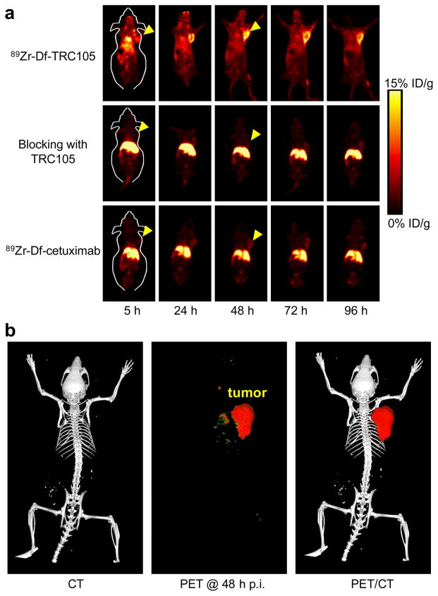
Small animal PET imaging of 4T1 tumor-bearing mice. a Serial coronal PET images at 5, 24, 48, 72, and 96 h post-injection of 89Zr-Df-TRC105, 2 mg of TRC105 before 89Zr-Df-TRC105 (i.e. blocking), or 89Zr-Df-cetuximab. Tumors are indicated by arrowheads. b Representative PET/CT images of 89Zr-Df-TRC105 in 4T1 tumor-bearing mice at 48 h post-injection.

