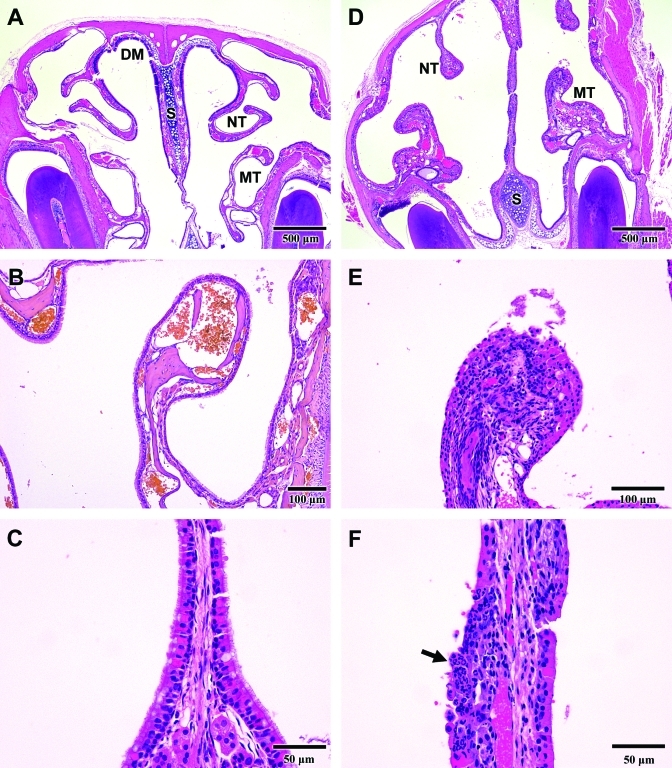
Figure 5.
Rhinitis. (A) Cross-section of a normal nasal cavity. Anatomic structures shown include the dorsal medial meatus (DM), nasoturbinate (NT), maxilloturbinate (MT), and nasal septum (S). (B) Normal maxilloturbinate. The surface is covered by a single layer of intact cuboidal or ciliated epithelial cells. (C) Normal respiratory epithelium lining the nasal septum is comprised of a uniform layer of pseudostratified, ciliated cells. (D) Nasal cavity from a mouse with rhinitis that was exposed to 120 ppm ammonia for 23 d. The nasoturbinates (NT) are blunted, and the maxilloturbinates (MT) and nasal septum (S) are thickened compared with normal structures (panel A). (E) A maxilloturbinate shows necrosis and sloughing of the surface epithelium with cellular debris in the lumen of the nasal cavity. The lamina propria contains a dense infiltration of inflammatory cells (compare with panel B). (F) Nasal septum from a mouse with rhinitis shows ulceration of the surface epithelium and dense infiltration with neutrophils (arrow).

