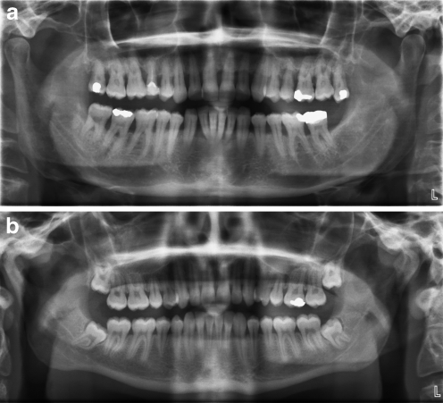
Fig. 3.
Periodontitis (radiographic appearance). a A 42-year-old man with type 2 diabetes and generalised severe periodontitis. There is extensive alveolar bone loss (generally 50–75% of the root length) affecting the entire dentition, with an irregular (uneven) pattern of bone loss. Some of the teeth have lost nearly all their supporting alveolar bone as a result of periodontitis progression, e.g. the upper molars (both right and left), and the four lower incisors, all of which are grossly mobile and which are retained in the oral cavity only by the soft tissue attachment (having lost 100% of their bone support). b A 21-year-old man with no periodontitis. Alveolar bone levels are normal, with the crest of the alveolar bone being in close proximity to the cemento-enamel junction (the boundary between the enamel crown and the root). Contrast with appearance in Fig. 3a

