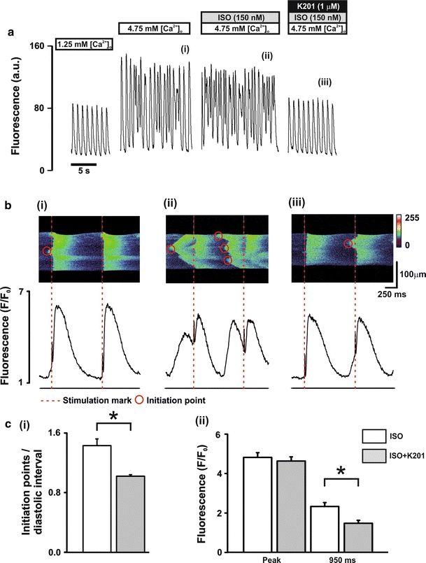
Fig. 4.

Characterisation of diastolic Ca2+ release events a perfusion protocol (upper) and typical records of Fluo-3 fluorescence (lower). b Typical line scan images (upper) and corresponding fluorescence profiles (lower) in the presence of (i) 4.75 mmol/L [Ca2+]o, (ii) 4.75 mmol/L [Ca2+]o + 150 nmol/L ISO and (iii) 4.75 mmol/L[Ca2+]o + 150 nmol/L ISO + 1.0 μmol/L K201; dashed lines (red) represent stimulus mark, circles represent wave initiation points. c mean ± SEM values of (i) number of wave initiation points/diastolic interval and (ii) fluorescence signals measured at the peak and at 950 ms post stimulation. White bars, ISO: 4.75 mmol/L [Ca2+]o + 150 nmol/L ISO; Grey bars, ISO + K201: 4.75 mmol/L [Ca2+]o + 150 nmol/L ISO + 1.0 μmol/L K201; n = 11, *P < 0.05
