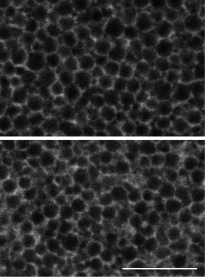
Figure 1.
High-magnification view of GFP–KDEL labeling in the cortex of immature oocytes. The top panel shows labeling in the animal half, and the bottom panel shows labeling in the vegetal half. On both sides, the ER has a network appearance, probably consisting of tubules and/or single (unstacked) cisternae. The pattern on the vegetal side has a small amount of patches. Cortical granules and other organelles are present in the dark spaces between the ER. Bar, 10 μm.

