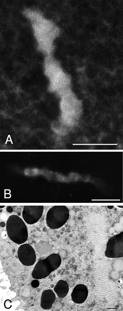
Figure 2.
Annulate lamellae in the vegetal half of immature oocytes. (A) An example of a long, dense island of GFP–KDEL labeling. These are present ∼5 μm in from the surface. Bar, 10 μm. (B) Immunofluorescence labeling with mAb 414 antibody to nuclear pores showing a similar structure as seen with GFP–KDEL labeling. Bar, 10 μm. (C) Thin-section electron micrograph in the vegetal cortex showing a long, narrow structure on the right side with the characteristic appearance of an annulate lamellae. The long, dense islands labeled by GFP–KDEL therefore correspond to annulate lamellae. Bar, 1 μm.

