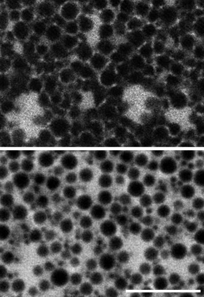
Figure 4.
Double labeling of GFP–KDEL (top panel) and cytosolic 3-kDa rhodamine dextran (bottom panel) in the vegetal cortex of mature oocytes. The cytosolic dextran penetrates into the cluster regions labeled by the GFP–KDEL. This indicates that the cluster is not a single swollen ER cisternae. This conclusion is consistent with the electron micrographs of ER clusters in Figure 5. The cortical granules and other large organelles in the cortex are seen in negative image in the dextran image, and the ER network outside the clusters is seen to run between these organelles. Bar, 10 μm.

