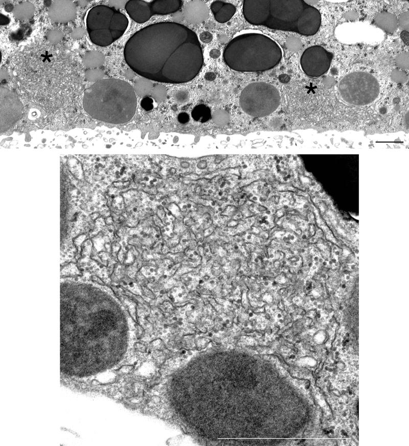
Figure 5.
Thin-section electron micrographs of ER clusters in the vegetal cortex of a mature oocyte. The top panel shows a low magnification view with two clusters that are denoted by black asterisks. The bottom panel is a high-magnification view of a cluster. The cluster consists of smooth-surfaced tubules and/or cisternae in a complicated three-dimensional arrangement. Bars, 10 μm.

