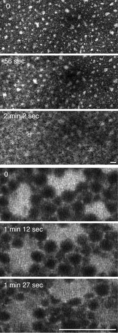
Figure 9.
Dispersal of GFP–KDEL-labeled ER clusters in the vegetal cortex during artificial activation. The egg was prick activated with a micro-needle, and then the egg was repositioned so that the vegetal cortex could be observed. The Ca2+ wave that is initiated by the prick activation takes 1–2 min to reach the region that is imaged. These two image sequences show the change in ER structure that occurs. The top three panels are a low-magnification sequence, and the bottom three panels show a higher-magnification view. Bars, 10 μm.

