
Figure 3.
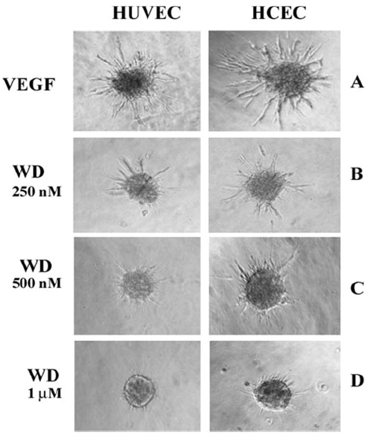
Withanolide D inhibited angiogenic sprouting in 3D-ECSA. Endothelial cell spheroids from HUVECs and HCECs were treated with 10 ng/mL VEGF in the presence and absence of different concentrations of withanolide D (WD). The HCEC-derived sprouts and HUVEC-derived sprouts were photographed after 36 and 24 hours, respectively.