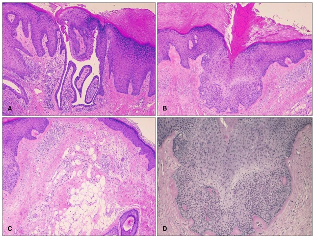
Fig. 3.
(A) Cystic invagination extended downward from the epidermis (H&E, ×40), which was diagnosed as a syringocystadenoma papilliferum. (B) There was a parakeratotic column overlying the trichoblastoma like-lesion, resembling the cornoid lamella of porokeratosis (H&E, ×40). (C) There were fat cells in upper dermis (H&E, ×40). (D) The basaloid epithelial proliferation showed PAS negative finding (PAS, ×100).

