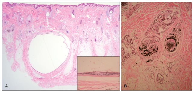
Fig. 2.
(A) The cyst was located within the lower dermis and subcutaneous fat (H&E, ×12.5). The folded cystic wall was lined by stratified squamous epithelium and contained flattened sebaceous gland cells (inset) (H&E, ×400). (B) Pigment casts were present in the hair papillae and peribulbar connective tissue (H&E, ×100).

