Figure 1.
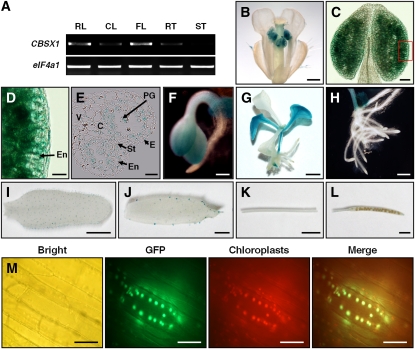
Expression Patterns and Subcellular Localization of CBSX1.
(A) RT-PCR analysis of CBSX1 expression. CL, cauline leaf; FL, flower; RL, rosette leaf; RT, root; ST, stem.
(B) to (L) Histochemical localization of GUS activity in ProCBSX1:GUS transgenic plant. From the 57 T2 transgenic lines, six individual lines were selected and examined for their GUS expression. C, connective tissue; E, epidermis; En, endothecium; PG, pollen grain; St, stomium; V, vascular region.
(B) Inflorescence cluster. Bar = 500 μm.
(C) Close-up view of anther. Bar = 50 μm.
(D) Magnified image of small box in (C). Bar = 20 μm.
(E) Transverse section of anther. Bar = 20 μm.
(F) Cotyledon of GUS-tagged line. Bar = 200 μm.
(G) Seedling stage of GUS-tagged line. Bar = 0.25 cm.
(H) Root of GUS-tagged line. Bar = 500 μm.
(I) Rosette leaf of GUS-tagged line. Bar = 0.5 cm.
(J) Cauline leaf of GUS-tagged line. Bar = 0.2 cm.
(K) Stem of GUS-tagged line. Bar = 0.5 cm.
(L) Silique of GUS-tagged line. Bar = 0.2 cm.
(M) Subcellular localization of CBSX1:smGFP protein. CBSX1:smGFP localized to chloroplasts. Bright, bright-field image; GFP, smGFP fluorescence; Chloroplasts, autofluorescence of chloroplasts; Merge, merged smGFP and chloroplast autofluorescence. The hypocotyl epidermal tissues of Pro35S:CBSX1:smGFP transgenic Arabidopsis seedlings (T2 generation) were observed under a fluorescence microscope. Bars = 20 μm.

