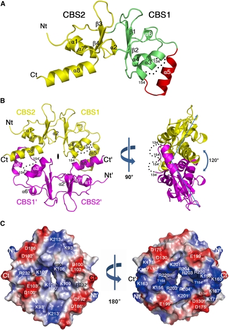
Figure 2.
Three-Dimensional Structure of CBSX2.
(A) Ribbon diagram showing the monomeric subunit structure of CBSX2. The first CBS domain (CBS1), the second CBS domain (CBS2), and an additional α-helical segment (α5) are colored green, yellow, and red, respectively. The secondary structural elements are sequentially labeled, and invisible residues (from 135 to 153) are indicated as black dots. The N and C termini of CBSX2 are labeled Nt and Ct, respectively.
(B) The overall structure of dimeric CBSX2 viewed along the twofold molecular symmetry axis (left). The two subunits are colored yellow and magenta. Prime (') is added to all labels of one subunit for clarity. Right: 90° rotation along the vertical axis, as indicated.
(C) Electrostatic potential surface of CBSX2 as viewed in (B), left. Positive and negative electrostatic potential are shown in blue and red, respectively. Right: 180° rotation along the vertical axis, as indicated.

