Figure 5.
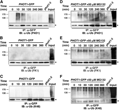
High-Intensity BL Stimulates Both Mono/Multi- and Polyubiquitination of phot1.
(A) When the 26S proteasome is functional, ubiquitinated phot1 is detected by an antibody that recognizes both mono/multi- and polyubiquitinated proteins. Total cellular proteins were prepared from phot1-5PHOT1-GFP seedlings that had been mock irradiated (0 min) or exposed to high-intensity unilateral BL (120 μmol m−2 s−1) for the indicated times. Total cellular proteins were also prepared from phot1-5 seedlings exposed to 120 min of high intensity BL as a negative control (lane 7). With the exception of an aliquot from the phot1-5PHOT1-GFP sample that was retained as a positive control for total cellular ubiquitination (Input, lane 8), all samples were subjected to immunoprecipitation (IP) with anti-GFP antibodies (lanes 1 to 7). Ubiquitinated proteins were detected by immunoblot (IB) analysis with P4D1 anti-Ub antibodies. Bottom panel shows an extracted portion of the same blot containing IgG heavy chain (~50 kD) from the α-GFP antibody (asterisk) as a loading control for IP samples.
(B) When the 26S proteasome is functional, no phot1 is detected by an antibody that recognizes only polyubiquitinated proteins. Replication of experiment in (A) except that ubiquitinated proteins were detected with the FK1 polyubiquitin-specific anti-Ub antibodies. Bottom panel represents a loading control as described in (A).
(C) When the 26S proteasome is functional, no phot1 is detected by an antibody that recognizes Lys-48–linked polyubiquitin chains. Replication of experiment in (A) except that ubiquitinated proteins were detected with anti-K48-Ub antibodies. Bottom panel represents a loading control as described in (A).
(D) When the 26S proteasome is inhibited, persistent phot1-Ub is detected. Replication of experiment in (A) except that seedlings were treated with 50 μM MG132 for 2 h prior to being mock irradiated or exposed to high-intensity unilateral BL. Bottom panel represents a loading control as described in (A).
(E) When the 26S proteasome is inhibited, persistent phot1 is detected by an antibody that recognizes only polyubiquitinated proteins. Replication of experiment in (B) except that seedlings were treated with 50 μM MG132 for 2 h prior to being mock irradiated or exposed to high-intensity unilateral BL. Bottom panel represents a loading control as described in (A).
(F) When the 26S proteasome is inhibited persistent phot1 is detected by an antibody that recognizes Lys-48–linked polyubiquitin chains. Replication of experiment in (C) except that seedlings were treated with 50 μM MG132 for 2 h prior to being mock irradiated or exposed to high-intensity unilateral BL. Bottom panel represents a loading control as described in (A).

