Figure 3.
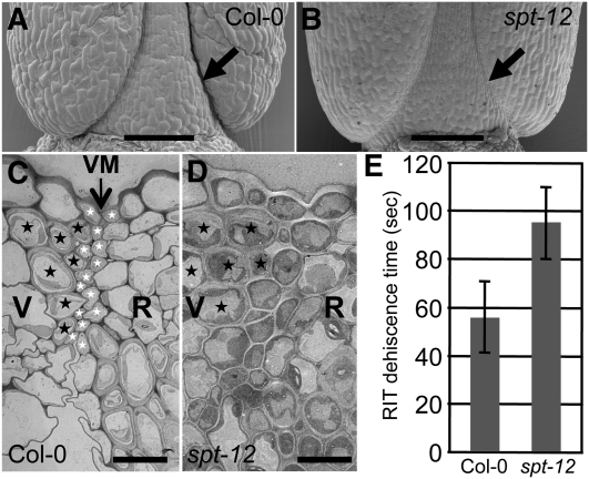
SPT Is Involved in Valve Margin Specification.
(A) and (B) Scanning electron microscopy of the base of Col-0 and spt-12 fruits at stage 18. Arrows indicate the dehiscence zone.
(C) and (D) Transmission electron micrographs of valve margin (VM) region in Col-0 and spt-12 siliques (stage 17b). Black and white stars indicate cells from the lignified and separation layers of the VM, respectively. Valves (V), VM, and replum (R) regions are indicated.
(E) Dehiscence assessment (Arabidopsis Random Impact Test) of Col-0 and spt-12. Values correspond to the time of shaking required to open 50% of dried siliques. Values are the average of at least three biological repeats (20 mature siliques for each) ± sd. The values are significantly different (Student’s t test P value = 0.02).
Bars in (A) and (B) = 100 μm; bars in (C) and (D) = 10 μm.

