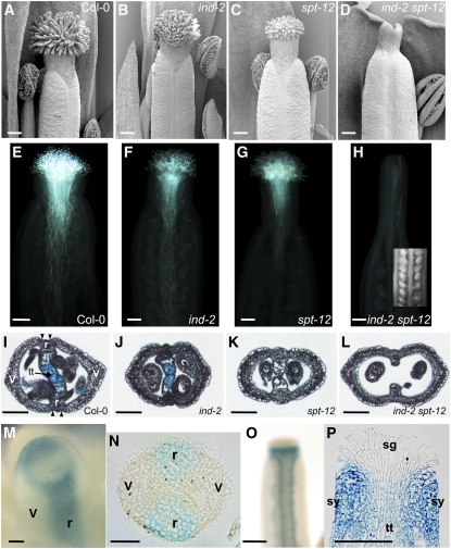
Figure 4.
IND and SPT Promote Formation of Marginal Tissues.
(A) to (D) Scanning electron microscopy images of gynoecia apical tissues at stage 13 in Col-0, ind-2, spt-12, and ind-2 spt-12.
(E) to (H) Pollen-tube growth in Col-0 (E), ind-2 (F), spt-12 (G), and ind-2 spt-12 (H). Inset in (H) is a light microscope image of ovules in an ind-2 spt-12 gynoecium.
(I) to (L) Cross sections of stage-13 ovaries in the wild type (Col-0) (I), ind-2 (J), spt-12 (K), and ind-2 spt-12 (L). Tissues are indicated in the wild-type section (I): v, valve; r, replum; tt, transmitting tract that has been stained with Alcian Blue.
(M) and (N) IND:IND:GUS expression in whole mount (M) and cross section (N) of stage-9 gynoecia. Presumptuous valves (v) and repla (r) are indicated.
(O) and (P) IND:IND:GUS expression in whole mount (O) and longitudinal section (P) of stage-12 gynoecia. Stigma (sg), style (sy), and transmitting tract (tt) are indicated in (P).
Bars in (A) to (H), (I) to (L), (O), and (P) = 100 μm; bars in (M) and (N) = 25 μm.

