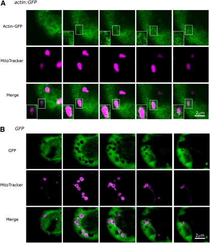
Figure 6.
The Existence of Filamentous Actin-GFP or Filamentous Bundle-Like Actin Patch in actin:GFP Mitochondria as Visualized by Confocal Microscopy.
(A) Five continuous sections of a protoplast cell from 12-d-old leaves of sterile transgenic actin:GFP tobacco plants are shown. Some actin-GFP fusion proteins (with no exogenously inserted mitochondrial targeting sequence) are apparently arranged in a filamentous bundle within mitochondria (stained with MitoTracker Red, CM-H2XRos, indicated in magenta). Inset boxes exhibit spots or filamentous bundle-like patches of merged actin-GFP and MitoTracker fluorescence (in white) in serial sections of a single mitochondrion.
(B) Five continuous optical sections of a protoplast cell from a leaf of a 12-d-old GFP transgenic tobacco plant, shown as a control for (A).

