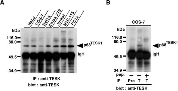
Figure 1.
(A) Immunoblot analysis of endogenous TESK1 protein expressed in various cell lines. Lysates prepared from approximately 5 × 10⁶ cells of each cell line were immunoprecipitated with anti-TESK1 antibody (TK-C21), run on SDS-PAGE, and immunoblotted with the same antibody. To detect endogenous TESK1 protein, the immunoblot membrane was exposed for 2 min in an ECL detection system. (B) Cell lysates of COS-7 cells were immunoprecipitated with an immunoglobulin G fraction of preimmune serum (Pre) or anti-TESK1 antibody (T) and immunoblotted with anti-TESK1 antibody. In the third lane, anti-TESK1 antibody was preincubated with excess amounts of antigenic C21 peptide. Positions of molecular size markers are indicated on the left. IgH, immunoglobulin heavy chain; IP, immunoprecipitation.

