Figure 2.
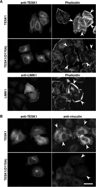
Stress fibers and focal adhesions induced by TESK1. (A) Actin organization induced by TESK1 and LIMK1. HeLa cells transfected with plasmids encoding TESK1, TESK1(D170A), or LIMK1 were stained with anti-TESK1 or anti-LIMK1 antibody (left) and rhodamine-labeled phalloidin for F-actin (right). (B) Focal adhesions induced by TESK1. HeLa cells transfected with plasmids encoding TESK1 or TESK1(D170A) were stained with anti-TESK1 (left) and anti-vinculin antibody (right). Arrowheads indicate cells expressing TESK1, TESK1(D170A), or LIMK1. Bar, 15 μm.

