Figure 9.
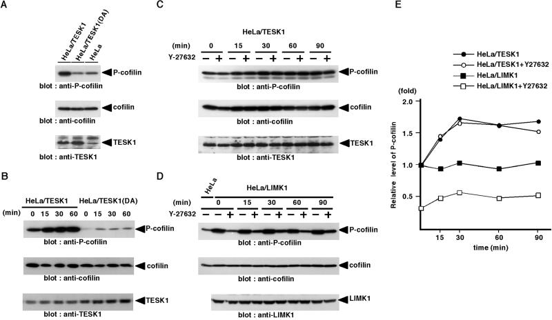
In vivo phosphorylation of cofilin by TESK1 is independent of Rho-ROCK signaling pathway. (A) HeLa cells stably expressing TESK1 (HeLa/TESK1) or TESK1(D170A) [HeLa/TESK1(DA)] were lysed, and the lysates were run on SDS-PAGE and analyzed by immunoblotting with anti-P-cofilin, anti-cofilin, and anti-TESK1 antibody. (B) HeLa/TESK1 or HeLa/TESK1(DA) cells were suspended and replated on fibronectin-coated dishes. At indicated times, cell lysates were prepared and analyzed by immunoblotting with anti-P-cofilin, anti-cofilin, and anti-TESK1 antibody. (C and D) HeLa/TESK1 or HeLa/LIMK1 cells were suspended, incubated for 30 min with or without 10 μM Y-27632, and replated on fibronectin-coated dishes. At indicated times, cell lysates were prepared and analyzed by immunoblotting with anti-P-cofilin, anti-cofilin, and anti-TESK1 (or anti-LIMK1) antibody. (E) Relative amounts of P-cofilin in HeLa/TESK1 or HeLa/LIMK1 cells treated with or without Y-27632 were plotted against the time after plating cells on fibronectin, with the amount of P-cofilin in HeLa/TESK1 or HeLa/LIMK1 cells without Y-27632 at zero time of plating taken as 1.0. Each value represents the mean of duplicate measurements.

