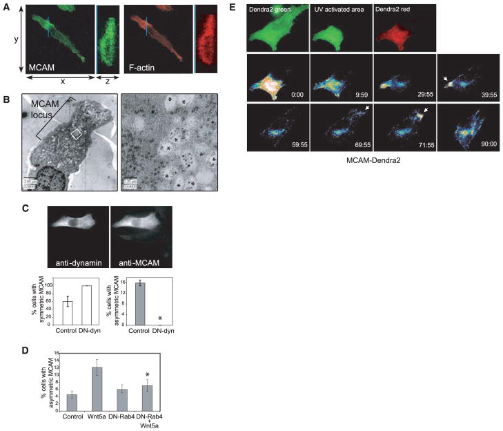
Fig. 4.
Formation and dynamic movement of the W-RAMP structure requires membrane internalization and endosome recycling and is associated with multivesicular bodies. (A) Confocal Z-series reconstructed in the region of the MCAM structure (blue line), showed that MCAM (MCAM-specific antibody, green) and F-actin (phalloidin, red) are localized to both cytosolic and membrane regions and are excluded from the nucleus. (B) Electron micrographs show accumulation of membrane vesicles associated with the W-RAMP structure. (Right) Higher magnification reveals that vesicles consist of multivesicular bodies (asterisks) interspersed among cytoskeletal filaments. (C) Cells expressing DN-dynamin, easily distinguished by their higher immunoreactivity, showed higher percentages with uniform symmetric MCAM localization and lower percentages with asymmetric MCAM localization (*P < 0.005, n = 3. Cells counted: control, 167; DN-dynamin, 203). (D) DN-Rab4 blocks the ability of Wnt5a to promote formation of W-RAMP structures, decreased significantly compared with Wnt5a alone (*P < 0.05, n = 4. Cells counted: control, 1019; Wnt5a, 1244; DN-Rab4, 1262; DN-Rab4+Wnt5a, 1034). (E) Red fluorescence from photoactivated MCAM-Dendra2 accumulates transiently in a perinuclear pool by 60 min, then at the opposite end of the cell by 71 min (arrow), followed by membrane retraction. Colors from lowest to highest intensity are black, blue, green, yellow, orange, red, and white.

