Figure 2.

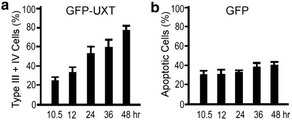
(a) Time dependent increase of type III and type IV cells exhibiting perinuclear aggregation of GFP-UXT. (b) Conventional apoptotic cells as in Fig. 1c expressing GFP alone. Data are the mean+SD of three independent experiments in which least 1,000 transfected type III plus IV cells or apoptotic cells were counted.
