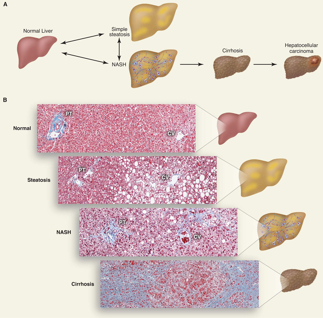
Fig. 1.
The disease spectrum of nonalcoholic fatty liver disease. (A) Schematic of progression of NAFLD. The accumulation of TG within lipid droplets in hepatocytes causes steatosis. Steatosis associated with inflammation, cell death, and fibrosis is referred to as NASH, which can progress to cirrhosis. Individuals with cirrhosis have an increased risk of hepatocellular carcinoma. (B) Histological sections illustrating normal liver, steatosis, NASH, and cirrhosis. Collagen fibers are stained blue with Masson’s trichrome stain. The portal triad (PT), which consists of the hepatic artery, portal vein, and bile duct, and the central vein (CV) are shown.

