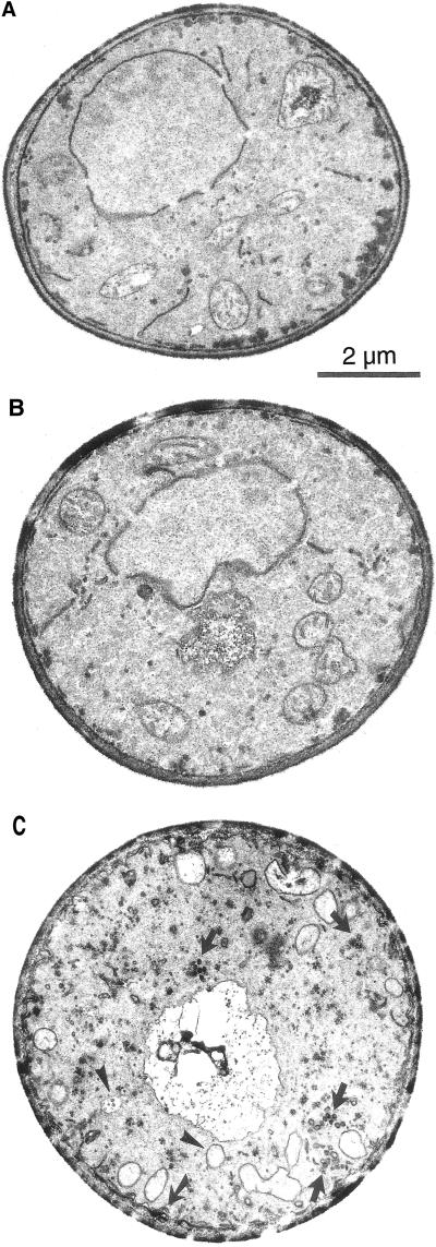
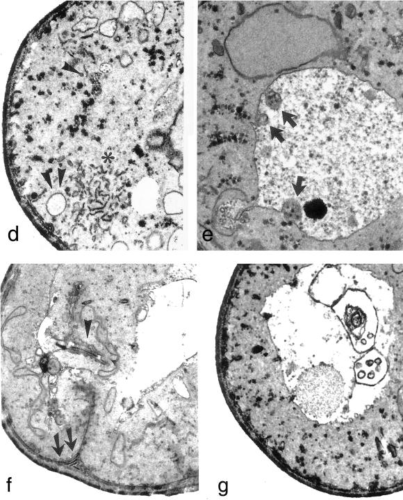
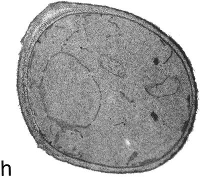
Figure 9.
(cont).Acb1p-depleted cells exhibit severe membrane alterations. Wild-type and Acb1p-depleted cells were grown in galactose (a and b, respectively) and in glucose media (h and c–g, respectively) and were prepared for electron microscopy. When grown on glucose, Acb1p-depleted cells accumulate vesicles of variable sizes (c, arrow), autophagocytotic bodies (c and d, arrowheads), randomly organized dense membrane areas (d, star), vacuolar inclusions of vesicles containing autophagocytotic like bodies (e, arrows), large invaginations of plasma membrane and accumulation of membrane material in the cytosol (f, arrowhead), and accumulation of membrane material in the vacuole (g).


