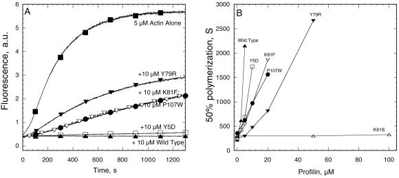
Figure 6.
Effect of profilins on spontaneous polymerization of 5 μM Mg-actin with 0.1 μM pyrene-labeled actin measured by pyrene fluorescence. (A) Time course of polymerization of actin alone (▪) and with wild-type profilin (▴), poly-l-proline binding mutant Y5D (□), and actin binding mutants Y79R (▾), K81F (▿), and P107W (●). (B) Dependence of the time required for half maximal polymerization on the concentrations of various profilin mutants. Symbols are same as in A with the addition of K81E (▵) that does not inhibit actin polymerization at even 100 μM.

