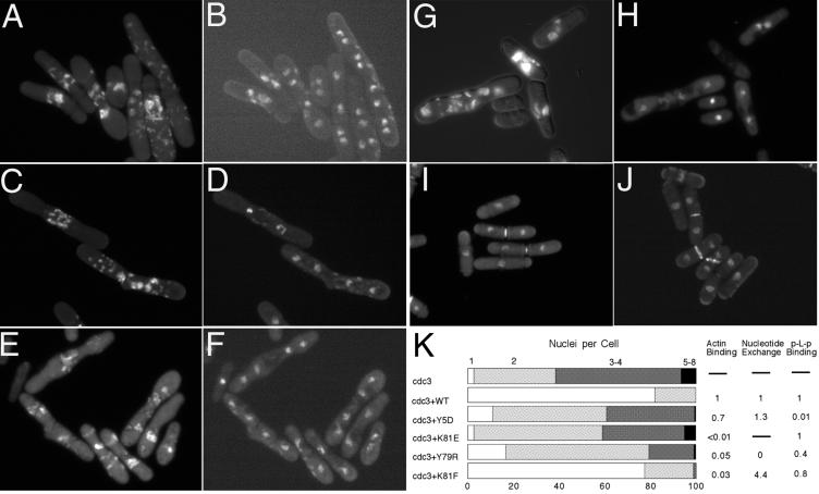
Figure 7.
Morphology of cdc3-124 ts strain expressing various profilins from pRep 81 plasmid under the control of the weakest nmt1 promoter after incubation for 8 h at 36°C in the absence of thiamine. Cells were fixed and stained with DAPI and Calcofluor. Calcofluor staining was recorded first (A, C, E, G). After bleaching of the Calcofluor, DAPI staining was recorded (B, D, F, H). (A and B) cdc3-124 without transformation. (C and D) Y5D. (E and F) K81E. (G and H) Y79R. Calcofluor and DAPI were recorded in the same time for wild-type profilin (I) and K81F (J). (K) Quantitation of nucleus number in comparison with biochemical data.

