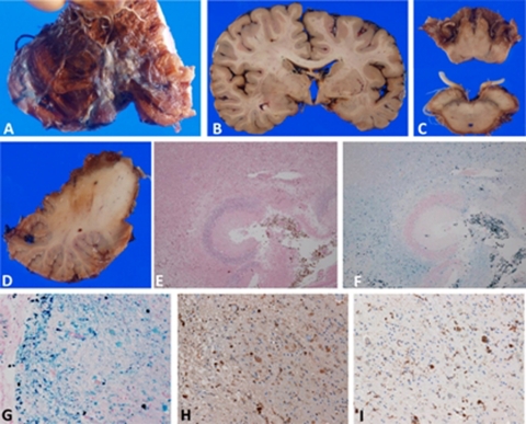
Figure 2.
Neuropathology of hemosiderosis. (A–D) Gross neuropathology. (A, D) Severe degeneration and brownish discolouration, indicating hemosiderosis, were observed at the anterior part of the cerebellum. Part of the left cerebellar hemisphere was removed and stored at –80°C for future analysis. (B, C) Hemosiderosis was present at the basal part of the cerebrum as well as the surface of the brainstem and cranial nerve roots. (E, F) Microscopic neuropathology. Severe necrotic lesions and hemosiderin deposits were observed in the cerebellar cortex (E, H&E stain; F, Berlin blue stain). (G) Severe iron deposits contained in macrophages were detected in the parahippocampus. (H) Some free iron deposits were also observed. Some ovoid bodies were present, which were also immunopositive for ferritin. (G, Berlin blue stain; H, ferritin immunohistochemistry; I, CD68 (for macrophages) immunohistochemistry) (E and F, 10× objective; G–I, 40× objective).

