Figure 3.
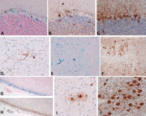
Microscopic neuropathology associated with tauopathy. (A–C) cerebellar cortex; (E and F) visual cortex; (G and H) parahippocampus. In areas with abundant iron deposits(A, G, E), numerous τ-immunopositive deposits were observed in the glial cells of adjacent sections (B, F, H). (D) τ-immunopositive neurons were present in the dentate nucleus of the cerebellum. (I) τ-immunopositive astrocytes were detected in the caudate nucleus. (J) Numerous τ-immunopositive neurofibrillary tangles were present in the locus coeruleus. (A and G, Berlin blue stain; B–E and H–J, immunohistochemistry using antibody (AT8) raised against phosphorylated tau protein.) (A–F, I, and J, 40× objective; G and H, 20× objective).

